
The creation of a new human being is one of nature’s most extraordinary processes. From a single cell to a fully formed baby, every stage of pregnancy unfolds with precision and wonder. Let’s journey through the key milestones that shape this remarkable journey.
The Prelude: Egg Maturation and Ovulation
Deep within a woman’s ovaries lie approximately half a million eggs, each nestled in a protective follicle. In every menstrual cycle, several follicles (and their eggs) begin to mature, but only one—the dominant follicle—grows to about 2 centimeters in size. As ovulation approaches, the fimbriated end of the fallopian tube (finger-like projections) moves to the ovary to “catch” the released egg.
The egg itself is layered for protection: an outer corona radiata and an inner zona pellucida. Both layers must be penetrated by a sperm to reach the egg’s nucleus, which holds 23 chromosomes—each carrying DNA, the blueprint for traits like eye color, body size, and organ function.
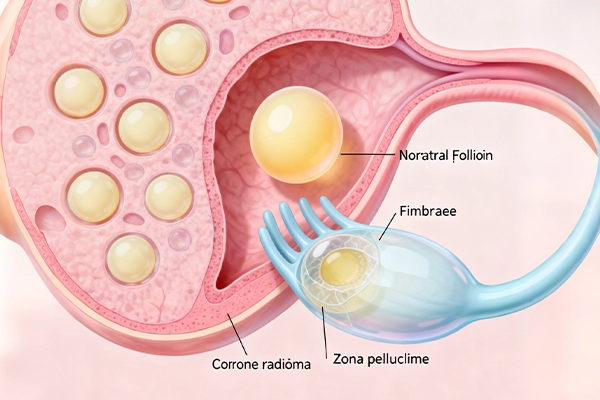
This image depicts the ovary with maturing follicles, the dominant follicle releasing an egg, and the fallopian tube’s fimbriae preparing to capture the egg. Labels highlight the corona radiata and zona pellucida of the egg.
Fertilization: The Union of Sperm and Egg
While the egg travels through the fallopian tube (propelled by cilia and muscle contractions at 3-4 mm per minute), thousands of sperm cells journey from the opposite direction. Guided by chemical signals from the egg, the sperm’s goal is to penetrate the egg’s protective layers.
When a sperm succeeds, its acrosome cap (a structure at the head) degrades, allowing it to enter the egg’s cytoplasm. This triggers two critical events:
- The zona pellucida hardens to block other sperm from entering.
- The egg completes its second maturation division, forming a female pronucleus and a polar body.
The sperm’s tail and head degenerate, and its nucleus expands to form a male pronucleus (with 23 chromosomes carrying the father’s DNA). The two pronuclei then replicate their DNA and align in a spindle apparatus—a step called syngamy. The result is a zygote, the first cell of the new human being.
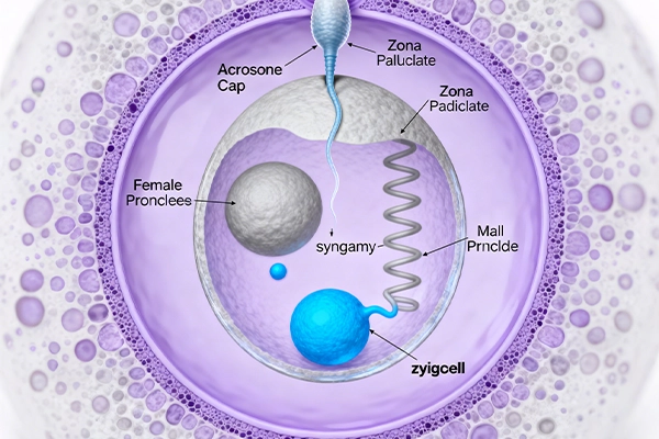
This cross-sectional image shows a sperm penetrating the egg’s corona radiata and zona pellucida. It highlights the formation of male and female pronuclei, followed by syngamy to form the zygote.
Embryonic Development: From Zygote to Blastocyst and Implantation
The zygote begins a process called cleavage, dividing repeatedly: 2 cells → 4 cells → 8 cells, and so on. By the 16-32 cell stage, it forms a mulberry-shaped structure called a morula, still enclosed by the zona pellucida (so cell size shrinks as cell number grows). Eventually, the morula develops into a blastocyst—a hollow, fluid-filled structure with two key parts:
- The embryoblast: The cluster of cells that will become the embryo.
- The trophoblast: The outer layer that will form the placenta and fetal membranes.
As the blastocyst reaches the uterus, the uterine lining (endometrium) is already thickened and nourished, ready for implantation. First, the blastocyst “hatches” from the zona pellucida, then burrows into the endometrium like a parasite. The trophoblast fuses with the uterine lining to form a nutrient-rich layer, while the embryoblast splits into the hypoblast and epiblast. Implantation may cause mild, harmless bleeding as the embryo burrows deeper.
Over time, the amniotic cavity (filled with protective amniotic fluid) and yolk sac (early nutrient source) form. The trophoblast expands to create the chorionic cavity and body stalk (which later becomes the umbilical cord).
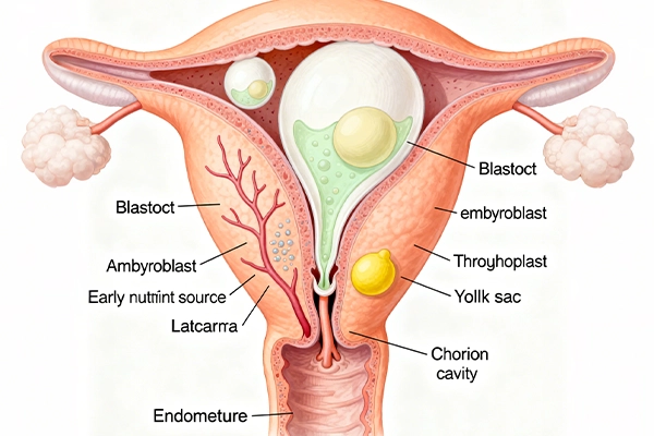
This side view of the uterus shows the blastocyst hatching from the zona pellucida, burrowing into the endometrium, and forming early structures like the embryoblast, trophoblast, amniotic cavity, and yolk sac. The uterine lining’s glands and blood vessels are labeled to show nutrient support.
Fetal Development and the Role of the Placenta
By the 3rd week, gastrulation occurs: the embryo’s primitive streak forms three germ layers (ectoderm, mesoderm, endoderm) that differentiate into all tissues and organs. The 3rd week also brings neurulation, where the neural groove closes to form the neural tube (the foundation of the brain and spinal cord).
By the 4th week, the embryo (now 4-5 mm long) has a beating heart, optic plaques (for eyes), and limb buds (upper buds first, then lower). It curves into a C-shape, and somites (cells that form bones, muscles, and skin) are visible. By the 8th week, the embryo is 23 mm long: fingers separate (toes follow later), and facial features like lens placodes (for eyes) and olfactory pits (for smell) form.
In the 9th week, embryogenesis shifts to fetogenesis. By the 12th week, the fetus has a human-like appearance: eyelids close, and skin is translucent. Over the next months:
- By 16 weeks, bones ossify (visible on ultrasound) and the body is covered in lanugo (downy hair) that traps vernix (a waxy coating protecting skin from amniotic fluid).
- By 18 weeks, the mother feels fetal movements, and the sucking reflex develops.
- By 28 weeks, eyes open, and head hair/eyelashes grow. Lanugo and vernix fade as the fetus gains mass.
The placenta (derived from the trophoblast) is vital for survival: it acts as a barrier between maternal and fetal blood (preventing mixing) while exchanging nutrients (carbohydrates, iron, vitamins) and oxygen from mother to fetus, and waste (carbon dioxide, urea) from fetus to mother. The umbilical cord connects the fetus to the placenta.
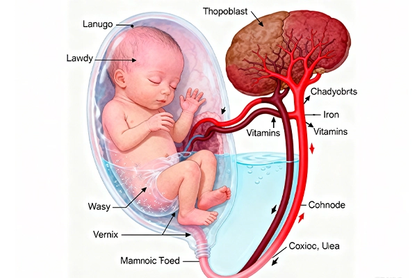
This image shows a mid-stage fetus in the uterus, connected to the placenta via the umbilical cord. Labels highlight key fetal features (limbs, closed eyelids, beating heart) and the placenta’s role in nutrient/waste exchange. Lanugo and vernix on the fetus’s skin are also depicted.
The Final Chapter: Birth
After about 38 weeks, the fetus is fully developed—typically 50 cm long and 3 kg in weight. Birth occurs within the next 2-4 weeks, marking the end of the pregnancy journey and the start of a new life.
From a tiny zygote to a breathing baby, every step of pregnancy is a testament to the marvels of human biology.

Good
Ggggg